

Archive position — measured, not model output
0 likes on Devpost
2,263 of the 7,850 archived projects have more likes, and 5,587 share exactly 0 — so this project's #3,021 place in the like-ranked listing is a tie-break inside that group, not a ranking.
Projects (log scale)
Likes on Devpost. ▲ marks this project's group.
Show the figures
| Likes | Projects | Share of archive |
|---|---|---|
| 0 | 5,587 | 71.2% |
| 1 | 1,758 | 22.4% |
| 2 | 284 | 3.6% |
| 3–4 | 132 | 1.7% |
| 5–9 | 75 | 1.0% |
| 10+ | 14 | 0.2% |
 User Stories BotA demo video on every pull request
Get a demo video on every new feature
GitHub App that watches your pull requests and records a narrated, captioned demo.
Save time on demo prep and make informed decisions faster with a video walkthrough of every new feature.
Executive Summary
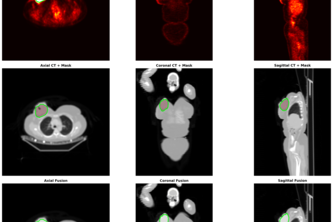
This is a self-reported research tool for breast tumour segmentation and radiomics from FDG PET/CT scans, built by one developer (Nitin Gupta) as part of an OpenAI hackathon submission. The project claims to automate the extraction of metabolic, shape, and radiomics-based measurements from breast cancer PET/CT data, including a novel metric called AMFIS. It is described as a Python-based workflow with GUI support, using DICOM/NIfTI inputs and leveraging tools like TotalSegmentator, PyRadiomics, and random-walker segmentation.
The author states that the tool aims to improve reproducibility and transparency in PET/CT tumour analysis for research use. No commercial or clinical deployment is evidenced; this appears to be a proof-of-concept or prototype-level system.
Key open question
Is there any evidence of real-world application, validation, or integration into clinical workflows beyond the author's own development?
What The Product Actually Is
The description states that this is a research workflow for breast tumour segmentation and radiomics from FDG PET/CT scans. It processes input in DICOM or NIfTI formats and outputs:
- Tumour segmentation
- Metabolic measurements (e.g., SUVmax, MTV, TLG)
- Shape-based metrics (e.g., asphericity, Dmax, 3D Feret diameter)
- Radiomics features (if PyRadiomics is installed)
- A novel metric called AMFIS (Asphericity-Metabolic Functional Inhomogeneity Score)
- Quality-control images for verification
The system uses Python and integrates libraries such as pyradiomics, simpleitk, scikit-image, numpy, matplotlib, and tkinter. It includes a GUI built with Tkinter.
Inference The tool is described as a research prototype, not a commercial product or clinical-grade software.
Positioning & Claim Evolution
The author positions the system as a way to turn routine FDG PET/CT studies into advanced tumour characterization tools. It is framed as:
- Improving reproducibility and transparency in PET/CT analysis
- Supporting research into disease progression, staging, and treatment response
- Providing outputs not typically available from standard viewing software
The system is described as a research workflow, not a clinical decision-support tool or commercial product.
Inference The positioning reflects an intent to support academic or research use, rather than direct clinical application or commercial adoption.
Target Customer & ICP
The description states that the tool is intended for researchers and clinicians working with PET/CT data in breast cancer studies. It is described as a tool for:
- Tumour characterization
- Disease progression and treatment response assessment
- Research into radiomics and metabolic heterogeneity
It is not stated whether it targets specific institutions, hospitals, or research groups.
Inference The ICP appears to be academic or research-oriented users, with no evidence of a defined commercial customer base.
Business Model & Pricing Evidence
There is no evidence in the description of any business model, pricing structure, or monetization strategy. The tool is described as a research prototype and not as a product for sale or licensing.
Inference No commercial business model is evident from the self-reported description.
Technical & Delivery Signals
The system is built in Python, using libraries such as:
pyradiomicssimpleitkscikit-imagenumpy,matplotlib,tkinter- Tools like
TotalSegmentatorandrandom-walkerfor segmentation - Support for DICOM/NIfTI input formats
It includes a Tkinter GUI for easier use, and supports optional PyRadiomics-style radiomics extraction.
The workflow is described as including:
- SUVbw conversion from PET metadata
- CT-guided breast-region localization
- Axillary and internal mammary node exclusion
- Random-walker segmentation with fallback logic
- AMFIS calculation
Inference The system is a custom-built research tool, not a commercial or scalable platform.
Traction & Maturity Signals
There is no evidence of any traction, adoption, or customer base. The project was submitted to a hackathon and is described as a prototype. No revenue, users, or clinical deployments are mentioned.
The author states that the next steps include:
- Improving robustness across datasets
- Adding lesion review/editing tools
- Validating metrics against clinical outcomes
- Packaging for easier testing in research environments
Inference The system is at early prototype stage, with no evidence of real-world use or maturity.
Competitive Context
There is no evidence of any competitive landscape or direct competitors mentioned. The description does not reference existing tools, platforms, or systems for PET/CT segmentation or radiomics in breast cancer.
Inference No competitive context is evident from the self-reported description.
Open Questions & Risks
- No commercial traction or adoption: The tool is described as a prototype with no evidence of real-world use.
- Single developer: The system was built by one person (Nitin Gupta), which raises questions about scalability, maintenance, and long-term support.
- No validation or clinical integration: No mention of clinical trials, peer-reviewed studies, or integration into existing workflows.
- Research-only focus: The tool is described for research use only, not for clinical deployment or commercial sale.
- Limited evidence of robustness: The author notes challenges in handling low SUV tumours and nodal interference, but no evidence of how these were resolved or tested.
Inference Risks include lack of commercial viability, scalability concerns, and limited real-world validation.
Questions the description leaves open
- What is the intended use case for this tool beyond research?
- Has the system been validated against clinical outcomes or peer-reviewed data?
- Are there any plans to integrate with existing clinical or research platforms?
- How does the tool handle edge cases or failures in segmentation?
- Is there any internal testing or feedback from clinicians or researchers?
- What is the roadmap for moving beyond prototype status?
Overall Assessment
The project is described as a research prototype built by one developer, with no evidence of commercial traction, clinical use, or business model. It is positioned for research purposes only.
Assessment Not evidenced as a viable investment or partnership opportunity at this stage. The tool lacks commercial signals and real-world validation. Any potential for future development would depend on further validation, integration, and scalability efforts.
User Stories BotA demo video on every pull request
Get a demo video on every new feature
GitHub App that watches your pull requests and records a narrated, captioned demo.
Save time on demo prep and make informed decisions faster with a video walkthrough of every new feature.
Source
Submitted to the OpenAI 2026 hackathon on Devpost. Project home on Devpost. Back to the full gallery on Devpost.
This competitive analysis was produced by top-founders.com (opens in a new tab)
Generated by a language model from this project's public Devpost page — its tagline, the author's own write-up and any built-with tags listed there — as captured in August 2026. Short quotations are the author's words. It is an opinion on the idea as a business, not on the people behind it, and it reflects the description as published then. If the project has moved on, request a correction below. The analysis is not independent research and contains no verified traction, revenue or customer data.
An independent guide by KFG International LLC — not affiliated with, endorsed by, or connected to Devpost or OpenAI.